Nuclear Magnetic Resonance NMR MRI
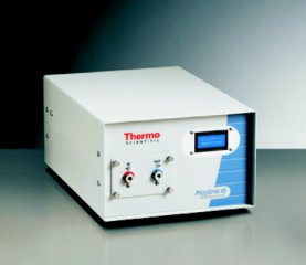
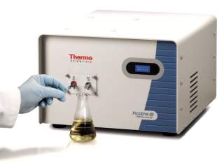
A nuclear magnetic resonance (NMR) spectrometer is an instrument based on physical phenomenon, in which nuclei in a magnetic field absorb and re-emit electromagnetic radiation. This energy is at a specific resonance frequency which depends on the strength of the magnetic field and the magnetic properties of the isotope of the atoms.
In a practical function of a NMR spectrometer, the frequency is similar to VHF and UHF television broadcasts (60–1000 MHz). NMR allows the observation of specific quantum mechanical magnetic properties of the atomic nucleus. Many scientific techniques exploit a NMR spectrometer to study molecular physics, crystals, and non-crystalline materials through.
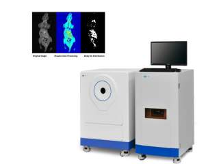
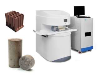
Magnetic Resonance Imaging (MRI) is also based on the same technology of a NMR. By measuring the relaxation time, T1 & T2 , of hydrogen-1 nuclei (1H), one can observe the disposition of water or hydrocarbons inside the tissue or sample. The disposition can be obtained from the image of the measured NMR signals. (Note: T1 and T2 is the time of hydrogen-1 nuclei from the excited state, returning to the stable state, where T1 is the spin-lattice relaxation time, longitudinal, and T2 is spin-spin relaxation time, transverse.)
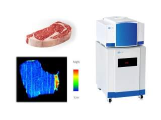
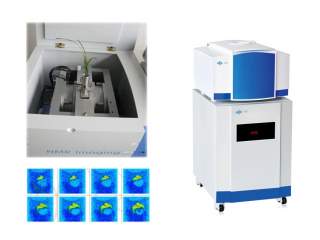
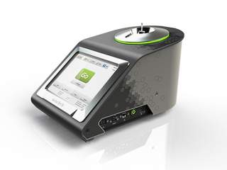
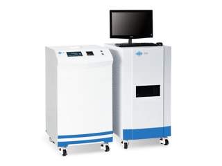
In recent years, Low Lield MRI becomes a very powerful tool in analyzing of biological samples, food samples, geological samples , fabrics samples, oil and petrochemical samples , because of the new technology and the large reducing of the cost and price. The best thing is, MRI is a non-destructive, fast method to analysis the state of the sample.